Tissue Imaging & Analysis Center (TIGA)
Prof. Dr. Niels Grabe
The Tissue Imaging & Analysis Center (TIGA) Center specializes in deep learning-based image analysis of digitized histological and cytological slides. This includes automatic segmentation, quality assurance, and assisted diagnostics. Our expanding AI classifier library, decentralized platform, and image database offer researchers and pathologists cloud-based access to whole slide images and results. The Center contributes to large epidemiological studies and small projects by generating valuable tissue and cytological data through automated scanners and advanced imaging systems.
Research Strategy
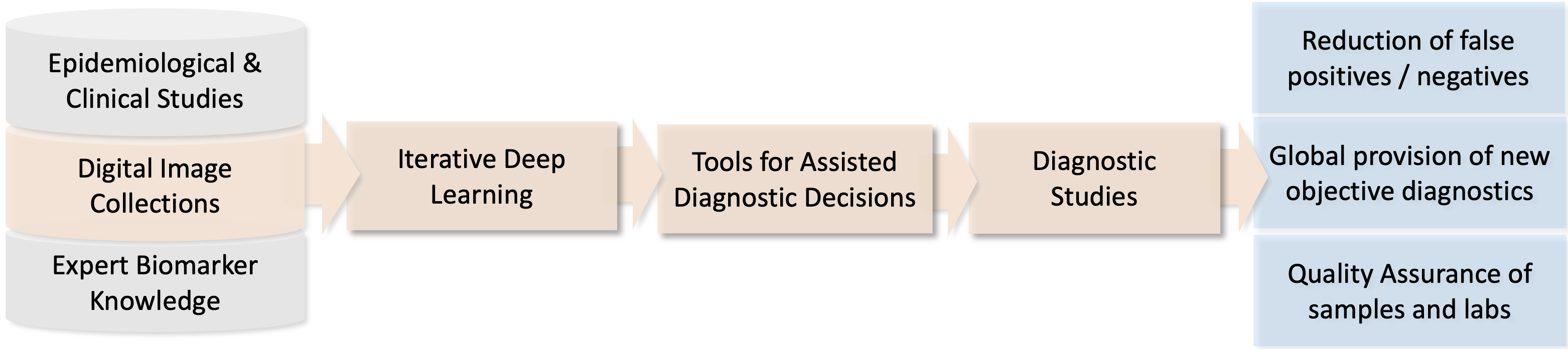
The TIGA Center’s core competence is deep learning-based image analysis of digitized histological and cytological slides. Depending on the application, the result can range from automatic segmentation to quality assurance of the prepared slide to assisted diagnostics. While work continues to expand the scope of the artificial intelligence (AI) classifier library, the range is also being expanded through the development of a decentralized deep learning platform and a digital image database. Whole slide images as well as the AI classifier results can be made available on a cloud application that can be used by researchers or as a diagnostics tool by pathologists (RUO).
In support of TIGA’s technology platform are automated microscopic scanners for whole slide imaging of glass slides. By integrating such imaging systems in a technical pipeline ranging from organotypic in vitro cell cultures to computational tissue modeling the TIGA generates a wealth of yet unexploited clinically highly relevant tissue and cytological data.
Tissue samples, unstained and stained histological slides and stained cytological slides can be processed. Tissue samples will be fixed, embedded and cut with the inhouse histological equipment and prepared for automated staining. Tissue sections are stained with in-house IHC staining machines. Stained slides are digitized with one of the four Hamamatsu NanoZoomer Digital Pathology (NDP) systems. The specimen can be scanned with either 20x or 40x magnification, yielding a high spatial resolution of 0.46 μm/pixel and 0.23 μm/pixel, respectively. The NDP systems detectors allow brightfield and fluorescence imaging of fixed tissue and cytology samples. The TIGA Center supports very large epidemiological studies such as with the US National Cancer Institute as well as smaller projects.

Project Leader

Prof. Dr. Niels Grabe
![]() +49 6221 54 51248
+49 6221 54 51248
![]() niels.grabe@bioquant.uni-heidelberg.de
niels.grabe@bioquant.uni-heidelberg.de
Selected Publications
Camryn M Cohen, Nicolas Wentzensen, Bernd Lahrmann, Diane Tokugawa, Nancy Poitras, Liam Bartels, Alexandra Krauthoff, Andreas Keil, Felipe Miranda, Philip E Castle, Thomas Lorey, Brad Hare, Teresa M Darragh, Niels Grabe, Megan A Clarke
Clin Infect Dis.2022 Oct 29;75(9):1565-1572
Nicolas Wentzensen, Bernd Lahrmann, Megan A Clarke, Walter Kinney, Diane Tokugawa, Nancy Poitras, Alex Locke, Liam Bartels, Alexandra Krauthoff, Joan Walker, Rosemary Zuna 7 , Kiranjit K Grewal, Patricia E Goldhoff 4 , Julie D Kingery, Philip E Castle, Mark Schiffman, Thomas S Lorey, Niels Grabe
J Natl Cancer Inst.2021 Jan 4;113(1):72-79
Jessica Kaufmann, Nicolas Wentzensen, Titus J Brinker, Niels Grabe
Oncotarget. 2019 Apr 2;10(26):2515-2529